Polymerase Chain Reaction (PCR) is a revolutionary molecular biology technique that amplifies specific DNA sequences exponentially.
Developed by Kary Mullis in 1983, PCR has become indispensable in pathology for diagnosing infectious diseases, genetic disorders, and cancers.
This article explores the principle, technique, variations, and clinical applications of PCR in modern pathology.
Principle of PCR
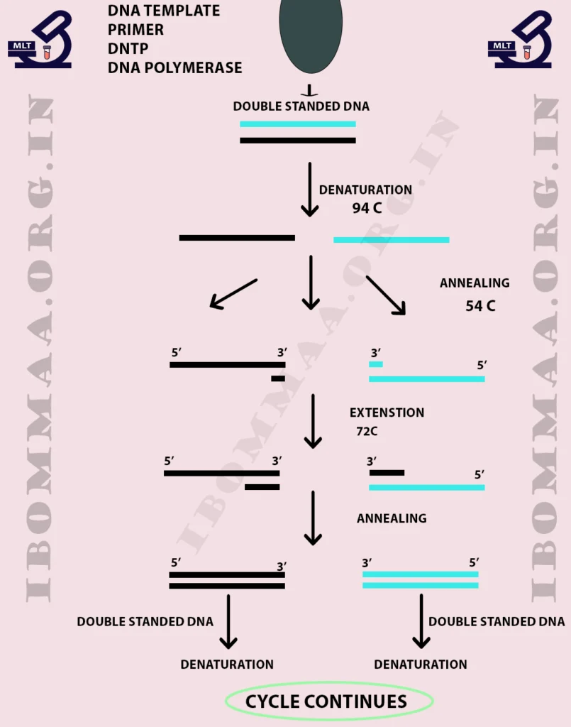
Polymerase Chain Reaction works by amplifying a target DNA sequence through repeated cycles of:
- Denaturation – DNA strands separate at 94–98°C
- Annealing – Primers bind to target sequences at 50–65°C
- Extension – DNA polymerase synthesizes new strands at 72°C
The process doubles DNA copies in each cycle, producing millions of copies in 20–40 cycles.
Key Components of PCR
| Component | Function |
|---|---|
| DNA Template | Target sequence to be amplified |
| Primers | Short oligonucleotides that bind to flanking regions |
| Taq Polymerase | Heat-stable enzyme synthesizes new DNA |
| dNTPs | Nucleotides (A, T, C, G) for DNA synthesis |
| Buffer Solution | Maintains optimal pH and Mg²⁺ concentration |
PCR Technique: Step-by-Step Procedure
1. DNA Extraction
- Isolate DNA from blood, tissue, or microbes.
- Purify to remove inhibitors (proteins, RNA).
2. Reaction Setup
- Mix template DNA, primers, Taq polymerase, dNTPs, and buffer.
- Load into a thermal cycler.
3. Thermal Cycling
| Step | Temperature | Duration | Purpose |
|---|---|---|---|
| Initial Denaturation | 95°C | 2–5 min | Unwinds DNA |
| Denaturation | 94–98°C | 20–30 sec | Separates strands |
| Annealing | 50–65°C | 20–40 sec | Primer binding |
| Extension | 72°C | 30–60 sec | DNA synthesis |
| Final Extension | 72°C | 5–10 min | Completes synthesis |
4. Product Detection
- Gel electrophoresis (visualization under UV light).
- Real-time PCR (quantification using fluorescent probes).
Types of PCR Used in Pathology
| Type | Application |
|---|---|
| Real-time PCR (qPCR) | Quantifies viral load (HIV, HBV, SARS-CoV-2) |
| Reverse Transcriptase PCR (RT-PCR) | Detects RNA viruses (Influenza, Dengue) |
| Multiplex PCR | Simultaneously detects multiple pathogens |
| Nested PCR | Increases sensitivity (low-abundance targets) |
| Digital PCR (dPCR) | Absolute quantification of rare mutations |
Applications in Pathology
1. Infectious Disease Diagnosis
- Detects bacteria (TB, H. pylori), viruses (HPV, HIV, COVID-19), and fungi (Candida, Aspergillus).
- Faster than culture methods (results in hours).
2. Cancer Genomics
- Identifies oncogenic mutations (EGFR, BRAF, KRAS).
- Monitors minimal residual disease (MRD) in leukemia.
3. Genetic Disorders
- Diagnoses sickle cell anemia, cystic fibrosis, thalassemia.
- Prenatal testing for Down syndrome (Trisomy 21).
4. Forensic Pathology
- DNA fingerprinting for identity verification.
- Detects microbial pathogens in autopsy samples.
Advantages of PCR
- High sensitivity (detects single DNA copies)
- Rapid results (within hours)
- Automated & scalable (high-throughput testing)
- Versatile (works on degraded samples)
Limitations
- Contamination risk (false positives)
- Requires precise primer design
- Cannot distinguish live vs. dead organisms
Conclusion
PCR is a cornerstone of molecular pathology, enabling rapid, accurate diagnosis of infections, cancers, and genetic diseases. With advancements like qPCR and digital PCR, its role continues to expand in precision medicine.
For pathologists and lab technicians, mastering PCR techniques ensures improved diagnostic accuracy and patient care.