Developed by Dr. George Papanicolaou, the Papanicolaou’s (PAP) stain is the gold standard in cytopathology, particularly for cervical cancer screening (Pap smears) and non-gynecologic exfoliative cytology. This polychromatic stain offers exceptional:
- Nuclear detail visualization (critical for dysplasia detection)
- Cytoplasmic differentiation (keratin vs. non-keratinized cells)
- Transparency (enables multilayer cell assessment)
Principle of Papanicolaou’s Stain
PAP stain combines regressive nuclear staining (hematoxylin) with progressive cytoplasmic counterstains (Orange G, EA-36) to highlight cellular features:
Key Components
| Reagent | Target | Color Produced |
|---|---|---|
| Harris hematoxylin | Nuclei | Deep blue |
| Orange G (OG-6) | Keratin/superficial cells | Orange-red |
| EA-36 (Eosin Azure) | Cytoplasm | Blue-green (non-keratinized) |
Step-by-Step Protocol – Papanicolaou’s Stain
1. Fixation & Rehydration
- Immediate 95% ethanol fixation (prevents air-drying artifacts)
- Gradual rehydration through:
- 70% ethanol (1 min)
- 50% ethanol (1 min)
- Distilled water (5 dips)
2. Nuclear Staining
- Harris hematoxylin (3.5 min)
- Acid alcohol differentiation (0.25% HCl, few dips)
- Bluing in lithium carbonate (1.5 min)
3. Cytoplasmic Staining
- Orange G (few dips): Highlights keratinized cells
- EA-36 (2 min): Stains non-keratinized cytoplasm
4. Dehydration & Clearing
- Absolute alcohol series (3 changes, 2 min each)
- Xylene (5 min till clear)
5. Mounting
- DPX mounting medium under coverslip
Interpretation Guide – Papanicolaou’s Stain
| Cellular Component | Normal Appearance | Abnormal Clues |
|---|---|---|
| Nuclei | Uniform dark blue | Hyperchromasia, irregular membranes (dysplasia) |
| Cytoplasm | Blue-green (non-keratinized) | Orange (keratinized) indicates maturation |
| Keratin | Bright orange | Excessive keratin (e.g., SCC) |
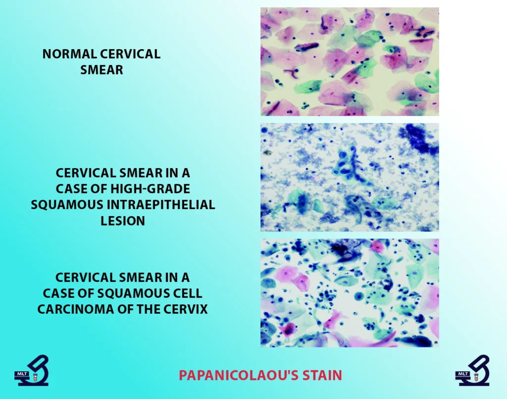
*Fig. 1: Normal cervical smear (400X) showing intermediate squamous cells with blue-green cytoplasm and uniform nuclei.*
Troubleshooting Papanicolaou’s Stain
| Issue | Cause | Solution |
|---|---|---|
| Pale nuclei | Exhausted hematoxylin | Filter or replace stain |
| Overstained cytoplasm | Prolonged EA exposure | Reduce staining time |
| Hazy background | Incomplete dehydration | Fresh alcohol changes |
Key Applications
- Cervical Cancer Screening
- Detects LSIL/HSIL (Fig. 2: HSIL with nuclear crowding)
- Identifies SCC (Fig. 3: Pleomorphic cells with orange keratin)
- Body Fluid Cytology
- Effusions (pleural/peritoneal)
- Urinary tract specimens
- FNA Specimens
- Thyroid, breast, and lymph node evaluations
Technical Pearls
- Fixation is critical: Process smears within 24 hours
- Stain freshness: Replace OG every 2 weeks, EA monthly
- Quality control: Run control slides with each batch
- LBC compatibility: Works with SurePath/ThinPrep systems
Conclusion
The PAP stain remains indispensable in cytopathology due to its unparalleled cellular detail preservation. Proper technique ensures accurate detection of premalignant changes (Fig. 2) and frank malignancies (Fig. 3). Modern liquid-based cytology has enhanced consistency, but the staining principles remain unchanged since Papanicolaou’s original work.
For optimal results: Maintain strict timing, fresh reagents, and proper bluing – the triad of perfect PAP stains.