Packed Cell Volume (PCV), also known as hematocrit, is a routine and vital blood test used in clinical pathology to determine the percentage of red blood cells (RBCs) in a given volume of blood.
This value reflects the oxygen-carrying capacity of blood and plays a crucial role in diagnosing conditions like anemia, dehydration, and polycythemia.
PCV is an essential parameter in complete blood count (CBC) tests and is commonly used in both hospital and laboratory settings.
Uses of PCV
- To diagnose anemia or polycythemia
- To monitor dehydration or blood loss
- For evaluating the response to treatment in hematological disorders
- To assist in calculating other indices like MCV (Mean Corpuscular Volume) and MCHC (Mean Corpuscular Hemoglobin Concentration)
- As part of pre-surgical evaluations and general health check-ups
Methods for Estimating PCV
There are two main techniques to measure Packed Cell Volume:
1. Microhematocrit Method (Micro Method)
Principle:
Blood is put into a capillary tube and centrifuged at high speed. The red cells settle at the bottom, and the percentage they occupy is measured as the PCV.
Materials:
- Heparinized capillary tubes (for capillary blood)
- Non-heparinized tubes (for venous blood with anticoagulant)
- Microhematocrit centrifuge
- Sealant (like clay)
- PCV reader or scale
Procedure:
- Fill 70–75% of the capillary tube with blood.
- Seal one end with clay.
- Centrifuge at 10,000–12,000 rpm for 5 minutes.
- Remove the tube and place it on a reader scale.
- Record the percentage of packed RBCs.
Interpretation:
- Normal PCV values:
- Men: 42–52%
- Women: 36–48%
- Children: 36–44%
- High PCV: Dehydration, polycythemia, high altitude
- Low PCV: Anemia, blood loss, overhydration
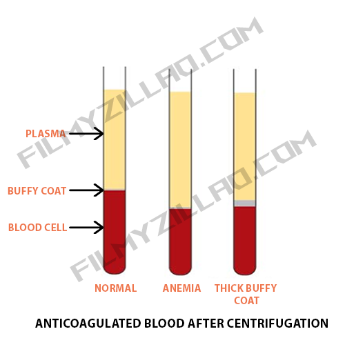
2. Wintrobe’s Method (Macro Method)
Principle:
Whole blood is filled into a Wintrobe tube and centrifuged. The height of the packed RBCs is read directly from the tube’s graduated scale.
Materials:
- Wintrobe tube (graduated up to 100 mm)
- Pasteur pipette or syringe
- Centrifuge
Procedure:
- Fill the Wintrobe tube with well-mixed anticoagulated blood up to the 100 mm mark.
- Centrifuge at 3,000 rpm for 30 minutes.
- Read the PCV directly from the height of the red cell column.
Comparison Table: Micro vs. Macro Method
| Feature | Micro Method | Macro Method (Wintrobe) |
|---|---|---|
| Sample volume | Very small (capillary blood) | Larger volume (venous blood) |
| Centrifugation time | 5 minutes | 30 minutes |
| Equipment used | Microhematocrit tube & centrifuge | Wintrobe tube & bench centrifuge |
| Speed of result | Faster | Slower |
| Accuracy | High | Slightly lower due to longer process |
| Common usage | Most commonly used method | Used for educational/demonstration |
| Buffy coat visibility | Limited | Better visibility |
Important Points
- Always mix blood gently to avoid clotting before testing.
- Use correct anticoagulants (e.g., EDTA) to prevent errors in results.
- Ensure accurate sealing of capillary tubes in the micro method.
- PCV results should always be interpreted alongside hemoglobin and RBC count.
- Errors in centrifugation time or speed may lead to inaccurate readings.
- Microhematocrit method is more suitable for small sample sizes and quick processing.
Conclusion
Packed Cell Volume is a simple yet powerful tool in clinical pathology for evaluating red blood cell concentration in blood.
Whether using the microhematocrit or Wintrobe’s macro method, accurate PCV measurement is essential for the diagnosis and management of various hematological and systemic conditions. The micro method is preferred in most modern labs due to its speed and reliability.