Haematoxylin and Eosin (H&E) Staining in Histopathology
Haematoxylin and Eosin (H&E) staining is the most widely used staining technique in histopathology. It provides detailed visualization of tissue structures, making it essential for diagnosing various diseases.
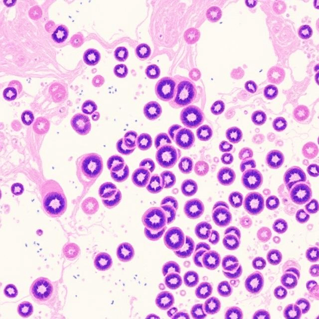
What Is H&E Staining?
H&E staining is a two-step process that uses two dyes:
- Haematoxylin: Stains the nuclei of cells blue or purple.
- Eosin: Stains the cytoplasm and extracellular components pink or red.
This combination creates contrast, allowing pathologists to differentiate between different tissue components.
Purpose of H&E Staining
- To highlight the overall structure of tissues.
- To identify abnormalities like inflammation, necrosis, or malignancies.
- To serve as a routine diagnostic tool in histopathology.
Procedure for H&E Staining
| Step | Process |
|---|---|
| 1. Fixation | Preserving tissue in a fixative (e.g., 10% formalin) to maintain structure. |
| 2. Tissue Processing | Embedding tissue in paraffin wax and sectioning it into thin slices. |
| 3. Deparaffinization | Removing wax using xylene and rehydrating tissue with alcohol and water. |
| 4. Hematoxylin Staining | Staining nuclei blue or purple by immersing slides in hematoxylin solution. |
| 5. Differentiation | Rinsing with acid alcohol to remove excess hematoxylin, leaving nuclei stained. |
| 6. Eosin Staining | Staining cytoplasm and extracellular components pink or red with eosin. |
| 7. Dehydration | Removing water from the tissue using alcohol and clearing with xylene. |
| 8. Mounting | Placing a coverslip over the tissue section using a mounting medium. |
Results of H&E Staining
| Tissue Component | Appearance |
|---|---|
| Nuclei | Blue or purple |
| Cytoplasm | Pink |
| Connective Tissue (e.g., Collagen) | Light pink |
| Red Blood Cells | Bright red |
Advantages of H&E Staining
- Cost-Effective: Simple and affordable for routine use.
- Diagnostic Utility: Provides clear differentiation of cell structures.
- Compatibility: Can be used alongside special stains or immunohistochemistry.
Limitations
- Does not identify specific proteins or pathogens.
- Requires additional staining techniques for specialized studies.
Conclusion
H&E staining is the cornerstone of histopathology, providing a clear view of tissue architecture and cellular details. Its simplicity and effectiveness make it indispensable for diagnosing various diseases.